
Drissi Lab
The Drissi Lab investigates the molecular pathways driving the differentiation and proliferation of high-grade pediatric brain tumors to identify and develop targeted therapies with the greatest potential impact for patients and their families. Our work encompasses a full translational research process as our projects range from in vitro work uncovering the role of epigenetic factors in these tumors to in vivo testing of experimental therapies in mouse models. Moreover, our lab has the privilege of collaborating with clinicians to bring therapies we study at the bench to bedside via clinical trials. It is through this relationship that we have the unique honor of deriving cell lines from the donation of patient tumor tissue, driving the development of better therapies by allowing us to advance our understanding of these poor-prognosis cancers.
Inside the Drissi Lab
Featured Publications
- Wang, Z., Kumar, A., Umaru, B. et al. Integrated multi-omics identifies lineage-dependent myeloid cells recruitment and the APP-CD74 axis as an immunoregulatory target in pediatric high-grade glioma. Acta Neuropathol 152, 39 (2026).
- Umaru B, Mishra DK, Kumar SS, Pang HH, Devine B, Khan K, Drissi R. BMI-1 modulation and trafficking during M phase in diffuse intrinsic pontine glioma. FEBS Open Bio. 2026 Jun;16(6):1213-1221. doi: 10.1002/2211-5463.70189. Epub 2025 Dec 27. PubMed PMID: 41455108; PubMed Central PMCID: PMC13238917.
- Mishra DK, Morris SM, Popovski D, Girard EJ, Bondoc A, Senthil Kumar S, Andrade AF, Zhu X, Yao F, Brusniak MY, Umaru B, Crotty EE, Brasel K, Pakiam F, Russo C, Zeinieh M, Biery MC, Coxon M, Conti H, Clarke M, Lu M, Rutka J, Llivichuzhca-Loja D, Konnikova L, Fouladi M, Jabado N, Huang A, Olson JM, Drissi R. Preclinical assessment of checkpoint blockade combined with DNA methyltransferase inhibition in high-risk pediatric brain tumors reveals limited therapeutic synergy. Neurooncol Adv. 2026 Jan-Dec;8(1):vdaf241. doi: 10.1093/noajnl/vdaf241. eCollection 2026 Jan-Dec. PubMed PMID: 41541220; PubMed Central PMCID: PMC12803780.
- Sengupta S, Senthil Kumar S, Bondra K, Sobo M, Mo X, Drissi R. Limitations of radiosensitization by direct telomerase inhibition to treat high-risk medulloblastoma. Front Oncol. 2023;13:1104670. doi: 10.3389/fonc.2023.1104670. eCollection 2023. PubMed PMID: 36741010; PubMed Central PMCID: PMC9891285.
- Liao Y, Luo Z, Deng Y, Zhang F, Rao R, Wang J, Xu L, Kumar SS, Sengupta S, DeWire-Schottmiller M, Berry K, Garrett M, Fouladi M, Drissi R, Lu QR. OLIG2 maintenance is not essential for diffuse intrinsic pontine glioma cell line growth but regulates tumor phenotypes. Neuro Oncol. 2021 Jul 1;23(7):1183-1196. PubMed PMID: 33539525; PubMed Central PMCID: PMC8661399.
Grants and Funding
FDA
“Phase II, Multi-arm, Molecularly-Guided Umbrella Trial for Children and Young Adults with High-Grade Glioma”
Role: Co-Investigator, PI: Fouladi
Pray Hope Believe Foundation
“Induced Telomere Damage and Cell Cycle Checkpoint Blockade: a Novel Approach for Radiosensitization and Immunomodulation to Treat Diffuse Midline Glioma and High-risk Medulloblastoma”.
Role: Principal Investigator
Department of Defense
“Molecularly-guided phase II umbrella trial for children, adolescents and young adults (CAYA) newly diagnosed with high-grade glioma (HGG), including diffuse midline glioma”
Role: Co-Investigator (PI: Maryam Fouladi)
MAIA Biotechnology, Inc.
“Induced Telomere damage and Radiation to Treat Poor-Prognosis Pediatric Brain Tumors”.
Role: PI
FDA
“A phase I and surgical cohort Study of PTC596 in newly diagnosed children with DIPG and high-grade gliomas”. Dr. Drissi is chairing the biology correlative studies (partial support of these studies).
Role: Co-Principal Investigator and Biology Chair
PTC Therapeutics, Inc.
“Preclinical testing of PTC596 and Ionizing Radiation to Treat DIPG and Pediatric GBM”
Role: Principal Investigator
Others
Cancer Free Kids Pediatric Cancer Research Alliance, National Center for Advancing Translational Sciences, The Cure Starts Now, The Lyla Nsouli Foundation
Join Our Team
The Drissi Lab is always growing. If you are interested in joining our team as a student, intern, research assistant/associate or post-doctoral fellow, please email us with a cover letter and a CV/Resume at Rachid.Drissi@NationwideChildrens.org.
Collaboration
Our lab collaborates with industry partners to advance research, drive innovation and translate scientific discoveries into real-world applications. This list highlights our partnerships with companies that support and extend the impact of our research.
- Company: Geron Corporation. Compound: Imetelstat. Target: Telomerase. Role: PI. Status: Studies completed.
- Company: Epizyme, Inc. Compounds: EP005687 and EPZ011989. Target EZH2. Role: PI. Status: Ongoing.
- Company: GSK. Compound: GSKJ4. Target: JMJD3. Role: PI. Status: ongoing.
- Company: Gilead Sciences, Inc. Compound: Momolotinib. Targets: JAK1, JAK2 and possibly ACVR1. Role: PI. Status: Completed.
- Company: PTC Therapeutics. Compounds: PTC-028 and PTC596. Target: BMI-1. Role: PI. Status: Completed.
- Company: PTC Therapeutics. Compounds: PTC596 in combination with radiation. Target: BMI-1. Role: PI. Status: Ongoing.
- Company: MAIA Biotechnology, Inc. Compound: THIO. Target: Telomeres. Role: PI. Status: Ongoing.
Patents
Our lab’s patents highlight innovative technologies, discoveries and research advancements developed through our work. This patent showcases our contributions to scientific innovation and the translation of research into practical applications.
Registered as Inventor at the U.S. Patent Office for the use of 6-thio-dG to treat therapy-resistant telomerase-positive pediatric brain tumors. International Application Number PCT/US20 19/023 596, International Publication Number WO 2019/183482 A1.
Featured Research Projects
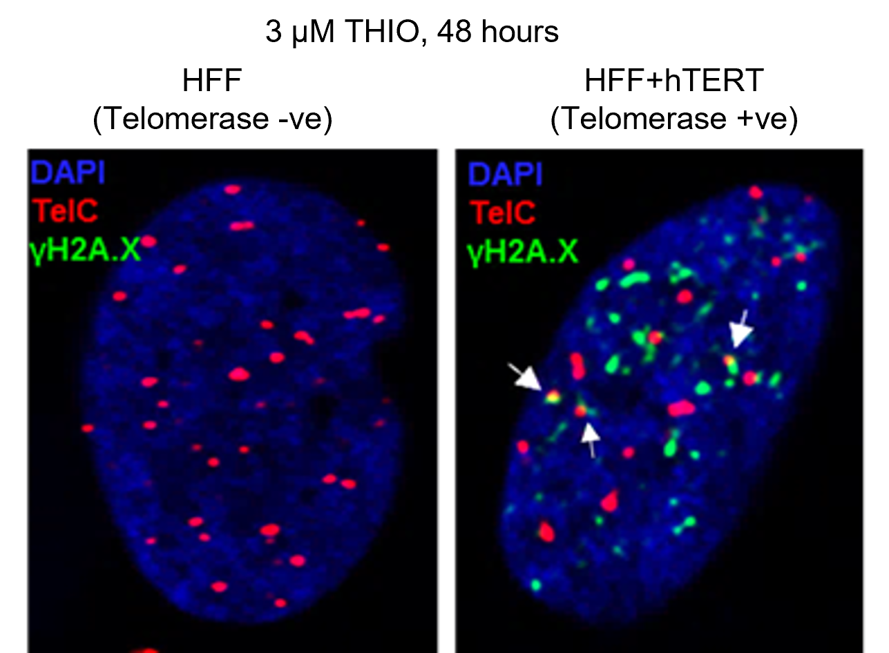
We used a novel strategy to induce telomere damage in telomerase-positive and therapy-resistant pediatric brain tumors. we showed that the telomerase-dependent incorporation of 6-thio-dG, a telomerase substrate precursor analogue, into telomeres leads to telomere dysfunction–induced foci (TIF) along with extensive genomic DNA damage. Our findings suggested that 6-thio-dG is a promising novel approach to treat therapy-resistant telomerase-positive pediatric brain tumors.
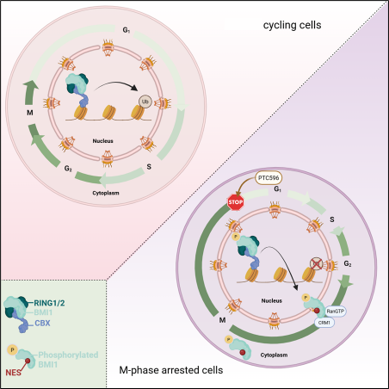
We identified BMI-1 as a therapeutic target in Diffuse Intrinsic Pontine Glioma (DIPG). The role of BMI-1 in DIPG was largely unknown. We showed that BMI-1 is highly expressed in tumor tissue samples of DIPG patients and in patient-derived cancer stem-like cells. Our data validated BMI-1 as a potential therapeutic target to treat children with DIPG. Furthermore, our findings indicated that BMI-1 modulation is associated with mitotic abnormalities, impaired DNA damage response, and cell death, supporting the combination of BMI-1 modulation and radiation as a promising novel therapy for children with DIPG.
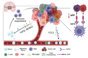
We identified mesenchymal-like tumor cells as strong drivers of monocyte recruitment in pediatric high-grade glioma and uncovered APP–CD74 as a candidate tumor–macrophage signaling axis. Reduced APP in DIPG tumors, together with the pro-inflammatory effects of APP on macrophages, suggests a potential role in shaping the immunosuppressive microenvironment.


