Single Left Ventricle Defects
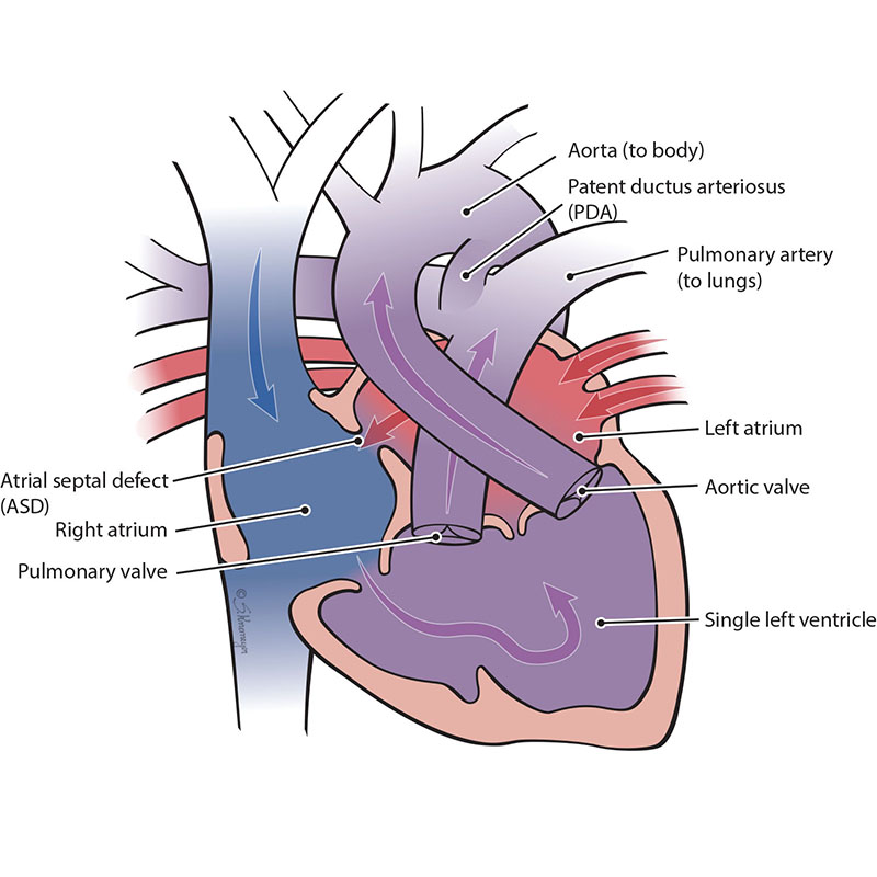
Double-inlet left ventricle is a condition when only the left lower pumping chamber (left ventricle) is fully grown (developed). The chamber that collects blood from the lungs (the left atrium) and the chamber that collects blood from the body (the right atrium) both send blood into a single left ventricle. The blood from the left ventricle is then pumped out of the heart through the pulmonary artery and aorta to the lungs and body at the same time. However, because all of the blood flowing through the heart mixes in the left ventricle, both red (oxygenated) blood and blue (deoxygenated) blood mixes together before going out to the body.
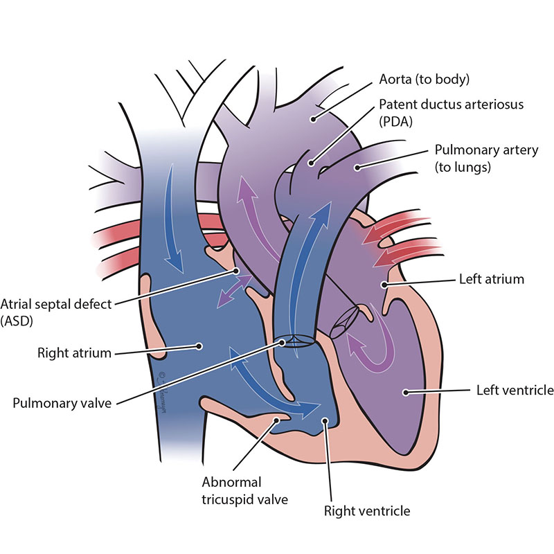
Ebstein’s anomaly is a heart defect where the tricuspid valve - the valve between the two right heart chambers (right atrium and right ventricle) - doesn’t work properly or is malformed. Blood may leak back through the valve, making your baby’s heart work harder. Ebstein’s anomaly may also lead to enlargement of the right side of the heart or heart failure. Many babies with severe Ebstein’s need single ventricle surgeries.
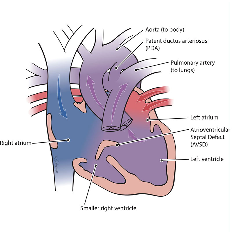
An atrioventricular septal defect (AVSD) is a heart defect where there are holes between both the top collecting chambers (atria) and bottom pumping chambers (ventricles). The valves between the atria and ventricles can be two separate valves or combined into one large valve. AVSD is also known as an atrioventricular canal defect (AVC or AVCD). In an unbalanced, left-dominant atrioventricular septal defect, the left ventricle is larger than the right ventricle. The difference in size between the ventricles can range from small to large. In severe cases, the right ventricle is too small to pump blood into the lungs.
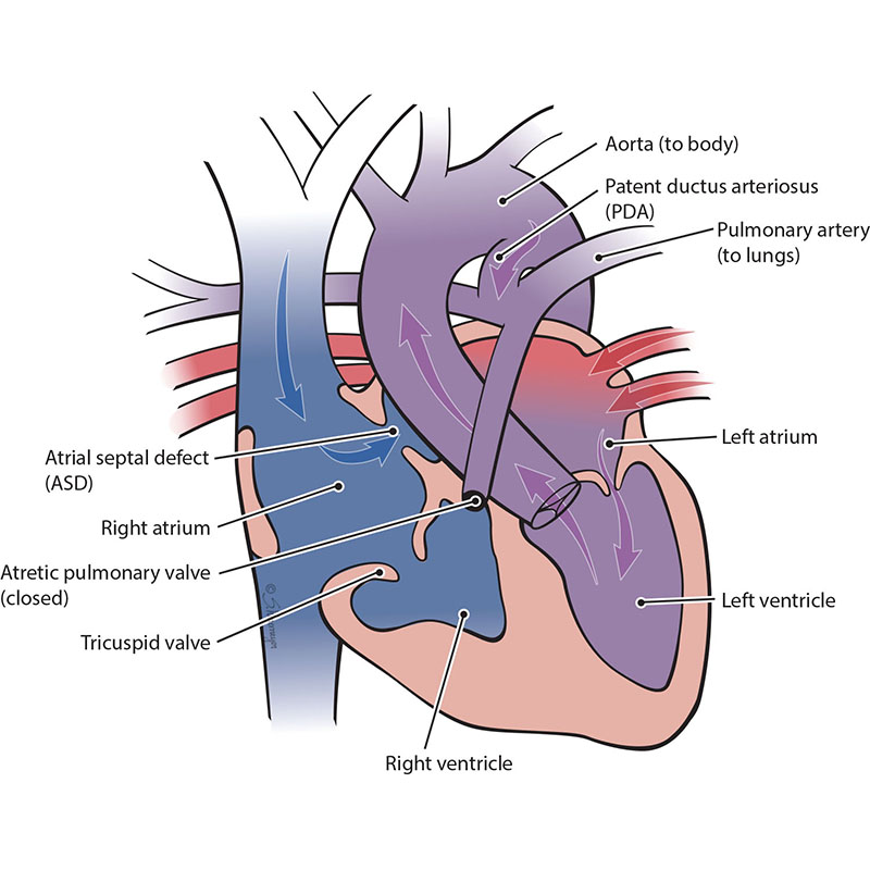
The pulmonary valve is a “door” that opens and closes between the right lower pumping chamber (the right ventricle) and the lungs. The condition known as pulmonary atresia with intact ventricular septum happens when the pulmonary valve stays closed and there is no hole between the bottom chambers of the heart. This means that blood cannot move from the right ventricle to the lungs like normal. When the right-sided chambers do not grow, this becomes a single ventricle defect. In this case, only the left ventricle is fully working.
The tricuspid valve is the “door” that opens and closes between the right upper collecting chamber (right atrium) and the right lower pumping chamber (the right ventricle). The condition known as tricuspid atresia is when the tricuspid valve does not grow (develop). This means blood cannot move from the right atrium to the right ventricle. When the right ventricle does not grow well, blood can only move into the right ventricle through a hole that is between the two pumping chambers. This hole is called a ventricular septal defect or VSD. Only the left ventricle develops to pump blood. This defect is sometimes called “hypoplastic right heart.” There are 2 main forms of tricuspid atresia depending on how the blood vessels connect, transposition of the great arteries or normally related great arteries.
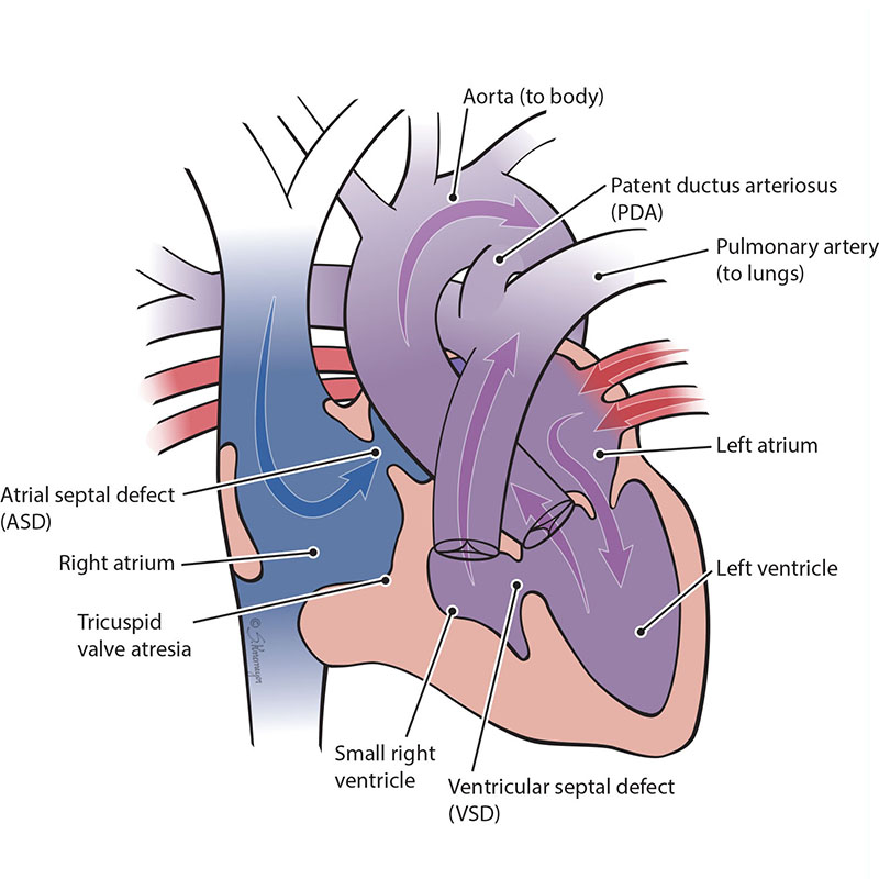
In Tricuspid Atresia with Normally Related Great Arteries, the aorta connects to the main left ventricle. The left ventricle pumps blood to the aorta (body) and through the hole (VSD) to the lungs (pulmonary).
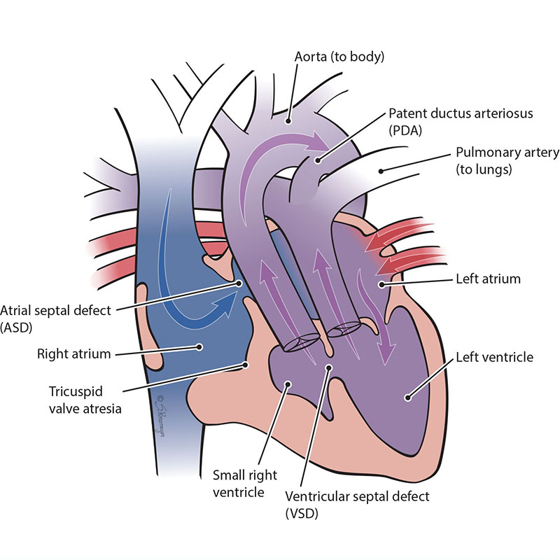
In Tricuspid Atresia with Transposition of the Great Arteries, the aorta connects to the small right ventricle. The body receives blood from the left ventricle through the VSD.