Congenitally Corrected Transposition of the Great Vessels (CCTGA)
In CCTGA, the receiving chambers (atria) are connected to their opposite pumping chambers (ventricles) because the ventricles are switched from their normal anatomic positions.
What Is CCTGA?
Congenitally corrected transposition of the great vessels (CCTGA; l-TGA) is an uncommon congenital heart abnormality, occurring in 0.5% of the population.
In CCTGA, the receiving chambers (atria) are connected to their opposite pumping chambers (ventricles) because the ventricles are switched from their normal anatomic positions. The aorta carries oxygen rich blood to the body, whereas the pulmonary artery carries oxygen poor blood.
In CCTGA:
- Normally situated aorta comes off of the inverted right ventricle and
- Normally situated pulmonary artery comes off of the inverted left ventricle
The blue blood from the body flows through the mitral valve to the left ventricle and is pumped out to the lungs to get oxygen. The oxygen-rich blood from the lungs flows through the tricuspid valve to the right ventricle and is pumped to the body. The right heart structures-tricuspid valve and right ventricle normally function at low pressure.
What Causes CCTGA?
In CCTGA, the receiving chambers (atria) are connected to their opposite pumping chambers (ventricles) because the ventricles are switched (inverted) from their normal anatomic positions.
What Are the Risk Factors?
Provided is a list of structural cardiac disorders commonly associated with CCTGA.
Pulmonary stenosis
This occurs in approximately 30-50% of CCTGA patients. There is muscular or valvular narrowing in the left ventricular outflow tract that results in diminished blood flow to the lungs. This can result in bluish discoloration (cyanosis) of:
- Skin
- Tongue
- Lips
- Nail beds
Ventricular septal defect (VSD)
This is an abnormal hole between the ventricles and occurs in 60-80% of patients with CCTGA.
Tricuspid Valvular Abnormalities
In CCTGA, the right ventricle is the "systemic ventricle", which pumps blood to the body. It must function at the body's blood pressure which is approximately four to five times higher than the pressure in the lungs.
Blood flows through the tricuspid valve, which is exposed to a high blood pressure, into the right ventricle. The tricuspid valve can be abnormally displaced in the lower right ventricular chamber which is called Ebstein anomaly. Aging patients with CCTGA can develop progressive leakiness.
Cardiac Rhythms Disturbances (Arrhythmias)
CCTGA can be associated with abnormalities in the heart’s electrical wiring. With advancing age, some patients have a significantly increased risk of developing atrioventricular (AV) block. The treatment for advanced AV block is to have a pacemaker implanted, which stimulates the heart, allowing it to beat fast enough. Symptoms of advanced AV block include:
- Lightheadedness
- Fainting
- Profound fatigue.
In patients with CCTGA, early heart beats (palpitations) and fast heart rhythms can arise from either the upper (atrial) or lower (ventricular) chambers.
Abnormalities in the specialized electrical tissues in the heart are common in CCTGA patients and can result in excessive slowing in the heart rate. Arrhythmias may require additional study of the heart's electrical wiring. Therapies needed to correct the heart rhythm can include:
- Medications
- Cardioversion/ablation
- Pacemaker
- Internal cardioverter defibrillator (ICD) implantation
What Are the Symptoms of CCTGA?
Although the heart has normal physiologic blood flow, anatomic abnormalities still exist. The right ventricle, which usually pumps blood to the low blood pressure lungs now pumps blood to the body (high pressure). The right ventricle handles the pressure load fairly well for many years but eventually begins to wear down, becomes dilated and, unfortunately, the right ventricle will weaken.
This weakening will be felt by patients as signs of heart failure:
- Shortness of breath
- Fatigue
- Chest pain
- Decreased exercise ability
Treatments with medicines are almost always helpful and can restore some of the heart function and improve symptoms. If the function of the right ventricle continues to deteriorate, a heart transplant may be an option.
A cardiac CT is performed in patients who are unable to safely undergo a cardiac MRI. Like a MRI, a CT also demonstrates anatomy of CCTGA. The image on the left is similar to the cardiac MR images above. The image on the right demonstrates the anatomy between the right ventricle (RV) and the aorta (Ao).
Heart failure symptoms include:
- Disproportionate
- Progressive fatigue
- Worsening shortness of breath with less and less activity
- Difficulty breathing during sleep.
The following diagnostic imaging studies are used to rack changes in the heart size and function:
Surgical Correction of CCTGA
Surgical corrections in CCTGA include:
- VSD repair using patches or sutures to close the defect
- Placement of a tube graft to overcome left ventricle outflow tract obstruction
- Tricuspid valve repair
Replacement for severe tricuspid valvular leakiness
What Is Life with CCTGA Like?
Pregnancy and Birth Control
Some congenital heart defects are passed down through families. For this reason, you may want to seek genetic counseling to determine your risk for having a child with a heart defect.
Pregnancy may increase certain health risks for women with heart defects, requiring close monitoring from experts in adult congenital heart disease and high risk maternal fetal clinicians. If you are trying to prevent pregnancy, you will need to select the lowest risk form of birth control.
In most cases, the cause of a congenital heart defect is not known. However, a child's risk for having a congenital heart defect increases if:
- A brother, sister, or parent has one
- Other genetic conditions are present (Down syndrome)
- The baby is born too early
Pregnant women or those who plan to become pregnant may be able to reduce their risk of having a baby with a congenital heart defect by taking steps to have a healthy pregnancy.
For a healthy pregnancy, avoid:
- Exposure to chemicals
- Exposure to certain diseases
- Use of certain medicines
- Cigarette smoking, tobacco, drugs, alcohol
- Exposure to extremely cold temperatures and places where you may not get enough oxygen
An unborn baby (fetus) has a higher chance of developing a congenital heart defect when the mother has diabetes or phenylketonuria.
Long term surveillance:
- Follow visit with experts in Adult Congenital Heart Disease
- Resting EKG
- Measurement of oxygen saturation
- 24 hour Holter monitoring
- Imaging studies
- Exercise stress testing to assess functional capacity
Other considerations:
Employment
Get an expert opinion from a cardiologist about your physical capabilities and risk for future heart problems as you explore career options. Also, recognize and be prepared for employers who may underestimate your physical capabilities. Even though no more than 10% of adults with congenital heart defects are considered disabled, some people may assume that all heart defects are serious and impair normal functioning.
Health Insurance
People with congenital heart defects often have difficulties when trying to change health insurance or when applying for new coverage. Research your options carefully before changing policies and find out whether you may qualify for state or federal assistance programs.
Use of Antibiotics
Most people with congenital heart defects have a lifelong increased risk for endocarditis and need to take antibiotics before having certain dental and surgical procedures.
Exercise
Patients with CCTGA can be quite active and participate in aerobic type activity:
- Walking
- Bike riding
- Jogging
- Swimming
Patients should always avoid heavy lifting or any activity that involves isometrics:
- Lifting
- Pulling
- Pushing
- Activities that cause grunting or straining
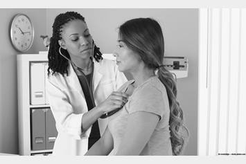
You need to talk to your doctor before getting involved in sports or exercising. You may need a cardiac stress test, sometimes done along with a type of echocardiogram, to measure how your heart responds to exercise.